|
Microscope Notes
Note: A compound microscope has 2 sets of lens. This kind of microscope is also called a light microscope as it requires a source of light to pass through the specimen.

| Microscope Parts |
Function |
| Arm |
Helps support the body tube that holds the lenses. The arm is used to carry microscope with the other hand holding the base. |
| Base |
Bottom part of the microscope that provides stability. |
| Ocular or Eyepiece |
The lens in the upper part of the microscope. |
| Body tube |
Holds the ocular lens and helps to reflect light towards the eye. |
| Nosepiece |
Holds the low and high power objective lens (4x,10x,40x). Can be rotated to change the magnification. |
| Objective lenses |
Located on the nosepiece. Holds the 3 to 4 objective lenses (4x,10x,40x, oil immersion lens). Only one objective lens can be used at a time. |
| Lower power objective lens |
Used to locate the specimen. |
| Higher power objective |
Allows for further magnification. |
| Stage |
The platform that supports the slide. |
| Stage clips |
Holds the slide in place on the stage. |
| Diaphragm |
Regulates the amount of light passing through the specimen. |
| Light source or illuminator |
Provides illumination for the specimen to be viewed. |
| Mechanical stage knobs |
Small knobs under the stage. Allows you to move the slide towards you, away from you, and side to side. |
| Coarse adjustment knob |
Located on the side of the arm. Moves the body tube up and down to approximate focusing. Only use when the low power objective lens is used. |
| Fine adjustment knob |
Located near the coarse adjustment knob. Used to sharpen the image during final focusing. |
Microscope Lighting
1. Under low power, the field of view is?
a. dark b. bright
2. If it’s too bright under low power, what should you do?
3. As you switch from low to high power, the field of view becomes?
a. darker with a decrease in diameter
b. brighter with an increase in diameter
4. If it’s too dark under high power, what should you do?
Answers:
1. Brighter
2. Partially close the diaphragm
3. Darker with a decrease in diameter
4. The diaphragm needs to be opened to allow in more light
Examining the Letter “e” on a slide
Note: When a wet mount of the letter “e” is made the following steps must be followed:
- Using a pair of scissors cut a small letter “e” from a piece of newspaper. Cut the smallest letter “e” you can find. Position the letter “e” on the center of a clean microscope slide.
- Using a dropper, place a drop of water on the piece of newspaper.
- Hold a clean cover slip in a vertical position (90 degree angle) next to the water. Make sure the bottom edge of the cover slip is in the drop of water on ones side. Lower the cover slip “rolling” it down at a 45 degree angle to avoid trapping air bubbles between the cover slip and the slide.
- Make sure the bottom of the slide is dry before you place it on the stage of your microscope.
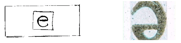
- Describe how the letter “e” will look under the microscope?
- While looking through the microscope, move the slide to the left, notice which way the letter “e” moved. Now move the slide to the right. Notice which way the letter “e” moved. Do the same with moving the slide away and towards you.
- Which knob should you never use while under a medium or high power objective? And Why?
Answers:
1. Image is reversed & inverted.
2. Moves to the opposite direction.
3. Coarse adjustment knob because the slide will crack.
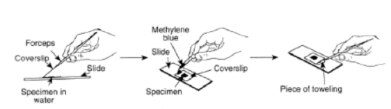
Making a wet mount of an onion cell
Note: When making a wet mount of an onion cell the following steps must be followed:
1. Place the specimen on a slide
2. Add a drop of water or stain.
3. Place a cover slip at an angle so that it touches the drop.
4. Lower the raised end of the cover slip slowly. The diagram below shows that as the cover slip is lowered, the drop of liquid moves to the right.

Staining an onion cell
Note: Staining an onion allows you to better observe structural parts of a plant cell. The staining reagents used are either iodine (Lugol’s solution) or methylene blue.

1. Prepare a wet mount (follow the above steps).
2. Place one drop of iodine alongside the coverslip.
3. Place a piece of paper towel at the edge of the opposite side of the coverslip. This will draw the stain under the coverslip and stain the cell.
Magnification
Power of the Eyepiece (ocular) X Power of the objective lens = Total Magnification of the Light Microscope
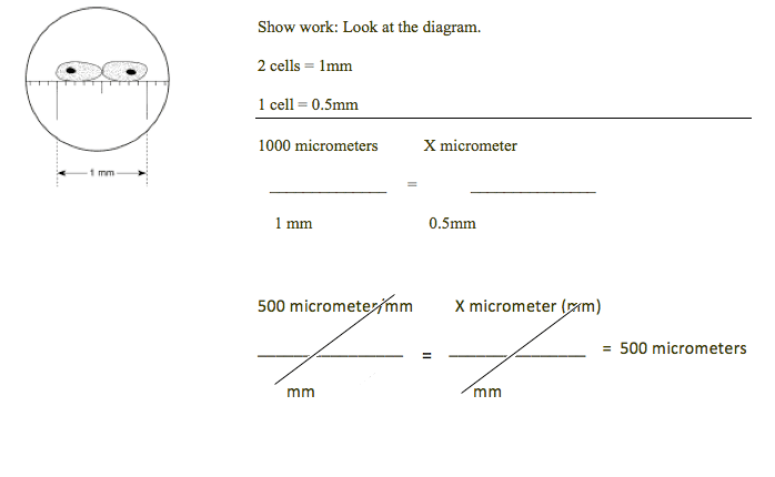
Measuring a specimen viewed under a microscope
- What is the average size of each cell in this field of view in micrometers?
Rule: 1,000 micrometers = 1 mm (millimeter)
2. The second image on the right is of the same organism as the first image on the left. What happened to form the image on the right?

Answer:
The student is viewing the same ameba on high power. The field of view gets smaller, which makes the ameba appear larger in this field.
|